The Chemistry
Section of the crime laboratory analyzes evidence for the presence of
Cannabis sativa L., kratom, intoxicating compounds and controlled substances as defined under the Illinois
Compiled Statutes, Chapter 720, Acts 550, 570, 643 and 690.
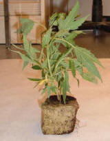
Cannabis
sativa L. plant
The laboratory
receives evidence for drug analysis in a variety of forms including
plant material, powders, liquids, tablets, capsules and paper. The most
common type of drug identified in the laboratory is Cannabis sativa L.
(also known as marijuana or pot). The next most frequently identified
drugs inlcude mixtures of heroin with fentanyl, cocaine and
methamphetamine. Some other drugs commonly
encountered in the laboratory include hydrocodone/acetaminophen mixtures
(Vicodin®);
benzodiazepines e.g. alprazolam (Xanax®), clonazepam (Klonopin®),
diazepam (Valium®); hallucinogens including lysergic acid
diethylamide (LSD), psilocin (found in “magic mushrooms”); ecstasy-type
drugs including 3,4-methylenedioxymethamphetamine (MDMA),
3,4-methylenedioxyamphetamine (MDA); anabolic steroids including
testosterone, stanozolol, nandrolone decanoate.
The drug analyst has
to first weigh the sample without packaging. This is achieved using an
electronic balance. Samples received in the laboratory can be as small
as a residue amount that is visible by eye, but not conducive to
weighing. They can also be very large. The laboratory has received on
occasion bales of cannabis (about the size of a microwave), and “bricks”
of cocaine (about the size of a hard-cover novel).
After weighing the
analyst then has to test the sample. At a minimum one preliminary test
and one confirmatory test has to be performed to be able to make
an identification.
Examples of
preliminary testing include color tests – wet chemical testing;
ultraviolet-visible spectrophotometry – measuring the absorbance of
ultraviolet light by the sample dissolved in a liquid; thin layer
chromatography – separating components of a sample on a chemically
coated glass plate; gas chromatography – volatilizing a liquid
preparation of the sample and separating components of the sample.
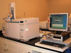
Gas
Chromatograph-Mass spectrometer (GC-MS)
Confirmatory tests
include mass spectrometry – breaking molecules into reproducible
fragments
and infrared spectrometry – measuring the degree of
transmission of infrared light through a sample. The data produced by
both of these methods is called a spectra and is akin to a chemical
fingerprint. For an unknown spectra to be identified as a controlled
substance it has to be compared to a known standard (purchased from a
chemical supplier, with a certificate of authenticity, that has been
verified in the laboratory by comparing its spectra to published data
before use in case work) run on the same instrument.
For the plant Cannabis
sativa L., a microscopical confirmatory test is performed.
This involves examining the unknown substance under a microscope and
observing for the presence of distinct morphological features.
On occasion the
section receives samples that despite thorough testing are not found to
contain a controlled substance. Some examples of commonly identified
non-controlled substances include baking soda, soap and vitamins.
CRIMINALISTICS
The Criminalistics Section evaluates and analyzes evidence for the presence of
fingerprints, palm prints and footprints. Any one of these types of
prints can be formed by the impression of the friction ridges (raised
portion) present on skin. The impression left can be made by the
natural secretions from sweat glands in friction ridge skin, referred to
as a latent print. A latent print is a crime scene print. Normally it
is invisible, but not always. Processing allows the print to be
visualized. Or they can be made by ink or other materials transferred
from the peaks of the friction ridge skin to a relatively smooth
surface, referred to as a patent print.
Two types of
evidence are typically submitted: lift-cards that have latent prints
taken from a crime-scene, and objects to be processed at the laboratory
for the presence of latent prints. The section receives a variety of
types of evidence for latent print processing. Some examples include:
paper, (e.g. checks, notebooks, letters, money); plastic, (e.g. plastic
bags, credit cards, bottles); glass, (e.g. bottles, mirrors); metal,
(e.g. guns, knives, cash registers).
The composition of
an object will determine which processing technique will be utilized to
develop latent prints. There are a variety of chemical and physical
methods that can be used including superglue fuming, luminescence
(laser), dye staining, powdering, ninhydrin and physical developer.
Many of these techniques can be used in tandem, for example, a plastic
bag would first be superglue fumed then processed with Rhodamime dye.
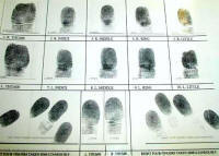
Fingerprint
Record
After the analyst
has processed the evidence to preserve and develop prints, the items are
photographed. The ridge detail observed in the photographs is then
examined to determine if it is of value for comparison. This evaluation
process is also conducted on submitted lift-cards. The first step is to
compare any prints that are of value to the victim’s record
fingerprints, or to anyone who had legitimate access to the crime
scene. Sixty to seventy percent of latent prints examined are
identified as belonging to the victim. The second step is to then
compare any remaining unidentified prints to the suspect’s fingerprint
record. The third step, if there are any remaining unidentified
fingerprints, is to enter them into the Automated Biometrics Identification System (ABIS)
(formely known as AFIS).
The Illinois State
State Police database contains over 3 million sets of fingerprints from arrestees,
job applicants, police officers and civil service employees in the State
of Illinois. The laboratory also has access to the Federal Bureau of
Investigation (FBI) Next Generation Identification (NGI) database.
A search of the ABIS
system will produce a list of persons whose fingerprint resembles the
entered print. The analyst will then obtain a copy of the original
fingerprint record to conduct the comparison. An identification is made
when the examiner determines that there are enough characteristics that
correspond in both the questioned and known print that allows the
examiner to conclude they were made by the same person.
FORENSIC BIOLOGY/DNA
Forensic Biology is
the area of the crime laboratory dedicated to finding and classifying
body fluids and biological substances from crime scenes. The
forensic biologist takes detailed notes, performs tests and collects and
preserves an appropriate amount of the evidence for subsequent testing.
Tests for body
fluids utilize chemical, enzymatic and microscopical techniques.
The body fluids most commonly tested for in the laboratory are blood,
saliva and semen. “Presumptive” tests indicate a body fluid may be
present. Preliminary chemical tests for body fluids usually
involve a color change. A small amount of blood – as little as a 1
to 1 million dilution – creates a fast blue/green color change with the
Tetramethylbenzidine (TMB) test. The chemical is reacting to the
iron molecule in red blood cells. This means that there is an
indication that blood is present, but there may be a few other
alternative explanations, even if they are less likely.
Presumptive tests
for saliva look for a chemical called amylase. Amylase is an
enzyme that begins breaking down starches in the mouth. A small
amount of the sample is placed in a gelatin that contains starch and is
allowed to incubate overnight. If amylase is present, it will
begin working its way outward from the center well where it was placed,
consuming the starch as it radiates out. Since the chemical iodine
will turn starch blue, an iodine solution is then added to the plate.
Where starch has been consumed, a clear circle is observed. The
diameter of the circle is proportional to the amount of amylase that is
present, indicating saliva.
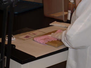
AP Press
Out on Clothing
Acid phosphatase, or
AP, is an enzyme that is found in many body fluids, but is at its
highest concentration in semen. This presumptive test also
requires a fast change to the color purple. The presence of semen, the
male reproductive fluid, can be identified by either the presence of
sperm cells or by the presence of relatively large amounts of Prostate
Specific Antigen, also known as PSA. Sperm are identified using a
stain and visually observing them through a microscope.
Usually, evidence is
not forwarded for DNA analysis unless a body fluid is identified.
Contact DNA from handled items is impossible to detect prior to
expensive and time-consuming DNA testing. There are four basic
steps in the DNA procedure: extraction, quantification,
amplification, and detection.
The extraction
procedure breaks open the cells containing DNA. It is important to have
the DNA in a liquid environment where it can flow and move, and thereby
interact with other chemicals that are used to test it. After the
DNA is released into the water, the sample is purified and concentrated
by removing excess water.
After the sample is
extracted, the analyst determines if there is any DNA in the liquid, and
if so, how much. The reaction of the extract is compared to the
reaction of a series of standards with known concentrations, called a
serial dilution. Based on this comparison, a rough estimate can be
made about the quantity of DNA present.
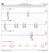
Electropherogram
The amplification
process, known as PCR (Polymerase Chain Reaction), targets specific
areas of the DNA and makes many copies of it. By looking at only
13 different locations in the DNA, enough characteristics can be
identified so that it becomes highly unlikely the DNA is from two
different randomly selected individuals.
In the final step,
detection of DNA products, only the STRs (Short Tandem Repeats) of
interest are observed because during amplification, a small chemical was
added to the DNA copy. This chemical will glow when a particular
kind of laser shines on it. The flash of light from the chemical
is captured by a digital camera and is displayed as a peak on print-outs
called “electropherograms”.
The DNA profile
obtained from the crime scene sample can then be compared to the DNA
profile from a known individual or it can be entered into a computer
database called Combined DNA Index System or
CODIS. This database
contains DNA profiles from convicted felons, from other crime scene
samples, from unidentified human remains and from other sources.